Following Circles: from the Lab to the Books to the Clinic, Exploring the Circle of Willis
Note: This post is based on the human dissection workshops I've been attending with Gil Hedley over the last couple of years.
A Word on Approaching Cranial Anatomy
Gil obliged my desire to see under the hood and so we removed the top of the cranium to reveal the mysteries inside! I find cranial anatomy utterly fascinating, it’s the terrain of the brain and it’s just so weird and beautiful. And, it’s kinda difficult to get a sense of.
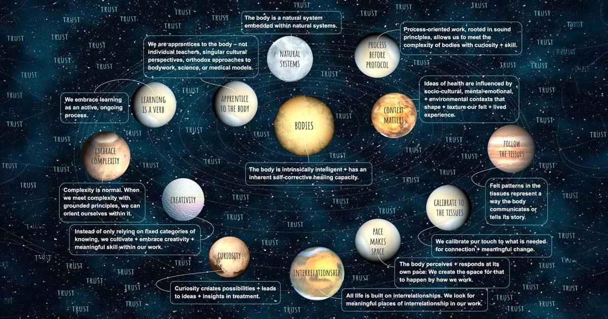
My approach to CST emphasizes the body, studying cranial anatomy is a way to support our hands connecting with this beguiling system and its infinite expressions. There are many ways to conceptualize CST, I like starting with the body, it keeps me grounded and connected to something tangible and allows the person receiving to let the work take them where it will. It’s not my role to direct their system to some intended place. Their system is wiser than I.
But why bother with cranial anatomy? Aren’t we just touching the bones, why does it all matter? These are good questions. It has been my experience that bodies can tell when hands and their imaginations are aligned with an image world of the tissues, the awareness of tissue relationships and realities seeps into our hands from our imagination and bodies can feel this. I think it creates a wordless trust. This empowers our touch in interesting ways and helps us to meet the always unique and unpredictable expressions of people’s bodies.
I am never far from the osteopathic principle of following the body, which means the body may have an idea about what it needs from the work that is different from the way I was taught. Do we, as clinicians, follow the wisdom of the body we are with in real time or follow the instruction of a teacher that is not present with this body? I will always choose the present body I am with over the instructions from a teacher… because that’s what I believe my teachers would do.
I have been a full-time clinician for over 25 years and I pay the most attention to what helps the folks on my table. This means I have expanded upon and moved away from prescriptive components of the craniosacral approach I was taught. As I have deepened my relationship with the anatomy of the system and continued exploring the underlying principles of the work within the crucible of clinical practice I feel more supported in the creativity that I feel is at the root of this work. These foci have greatly enhanced my clinical impact.
With this in mind, let’s look at some of the circulatory anatomy of the craniosacral system, it’s gorgeous!
Mind-Blown-Emoji - Skull Popped! 🤯
It’s so wild to have the saw come out and cut the cranium so we can peer into the mystery. In an unembalmed cadaver, the brain tissue itself is decomposed and pretty squishy and not super interesting. In an embalmed cadaver the brain tissue is intact and available for exploration. I have had the opportunity to study both.
On Gil’s website there are incredible cranial dissections. I highly recommend getting a subscription and watching the videos.
Every dissection contains its own teaching, it’s parallel to doing listening based bodywork. You might start with an idea and then the body has something much different it wants to do. So, we follow that and learn together.
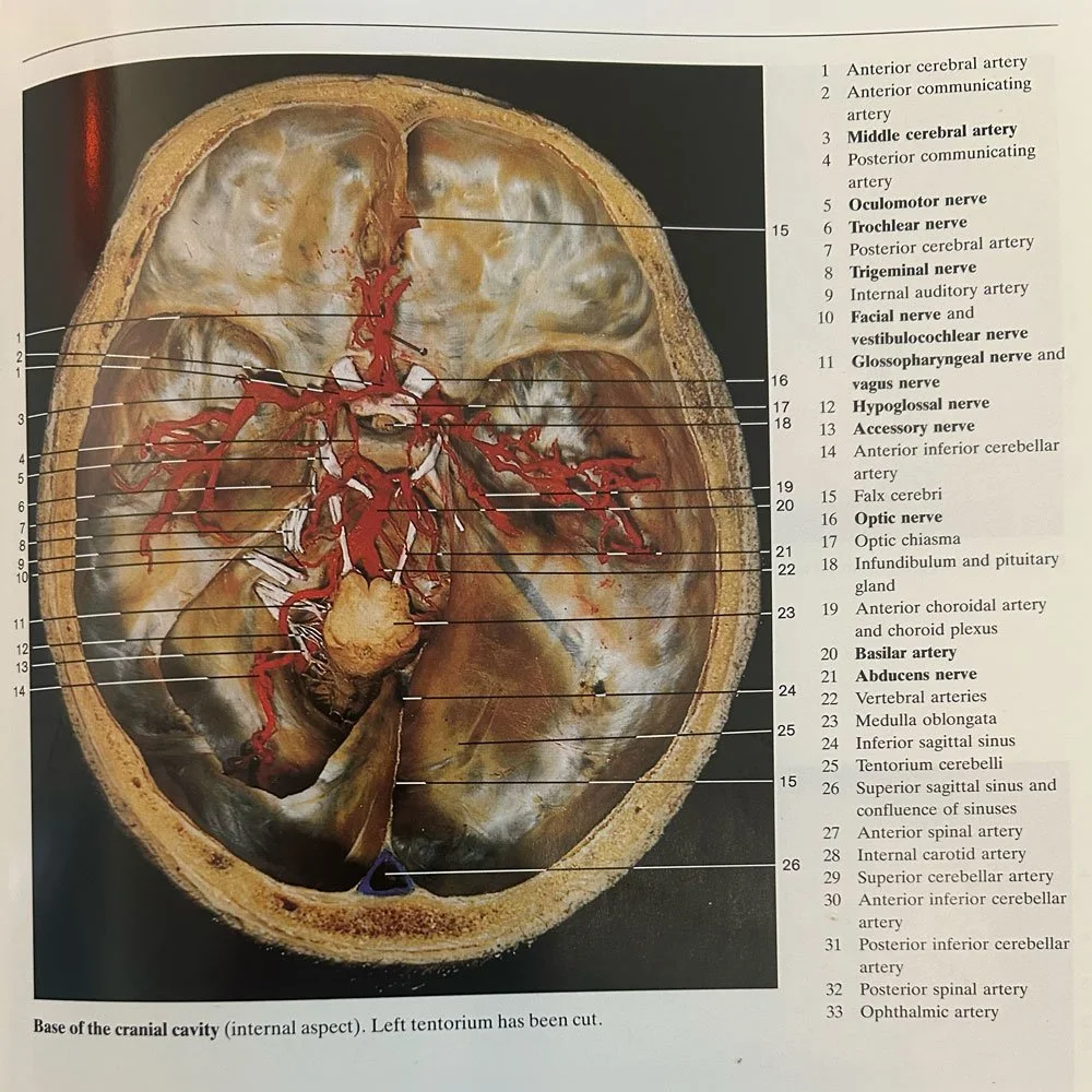
Color Atlas of Anatomy, 3rd Ed. (Rohen / Yokoshi)
This body offered a beautiful expression of the emergence of some of the structures we can see when the cerebrum is lifted away. The membrane system is left covering the surfaces of the temporal bones, the sphenoid and the frontal bone. Posteriorly, the membrane forms the tentorium, which covers the cerebellum and provides for separation between the upper and lower portions of the brain.(1) It’s a super important structure within our approach to CST. The picture below gives a great view of this.
On the left the tentorium is cut away and you can see the cerebellum. On the right it is left intact and you see how it forms a “tent” over the cerebellum. What sometimes is left out of the conversation is that the membrane is more than the tentorium. It covers the entire surface of the cranial base and is potentially involved with all of the structures we find there. In the image you can see the membrane totally contouring the multiple ridges and depressions of the temporal bones, sphenoid and the frontal bone. This membrane is contiguous with the entire membrane system and the cerebrum rests (and maybe floats a little due to some buoyancy offered by the fluid system within the cranium) on top of this membrane.
When I registered this it was a game changer for me and helped me reconceptualize my sense of how the membranes may be transmitting tension and forces from our work? This is an open question and a bit of a working hypothesis. I don’t think there are many facts when it comes to the hows and whys regarding the various ways that folks respond to CST. There is mystery here.
In this image, we have an utterly amazing view of the arteries of the brain. It’s just gorgeous. In the lab, we did not see this specific beauty! It was part of smushy stuff that didn’t hold form (I’m curious if these arteries were dyed?). But we could see antennae like projections of the arterial structures that form the circle of Willis or the cerebral arterial circle (preferred name). Seeing these little arterial projections was stunning. They form the arterial system for your brain, meaning it's the oxygen delivery pathway for everything our sweet little brains try to do in a day.
This potentially has implications for folks dealing with POTS and other forms of dysautonomia.
Seeing them inspired me to go back to the back and study this anatomy more. In the lab is a library of texts we regularly reference. As we iterate between the body and books we form a more and more dynamic impression of what and how bodies are. The truth is there is no one truth about bodies. Our understanding of them is best held as impressionistic so that we leave space for ongoing learning.
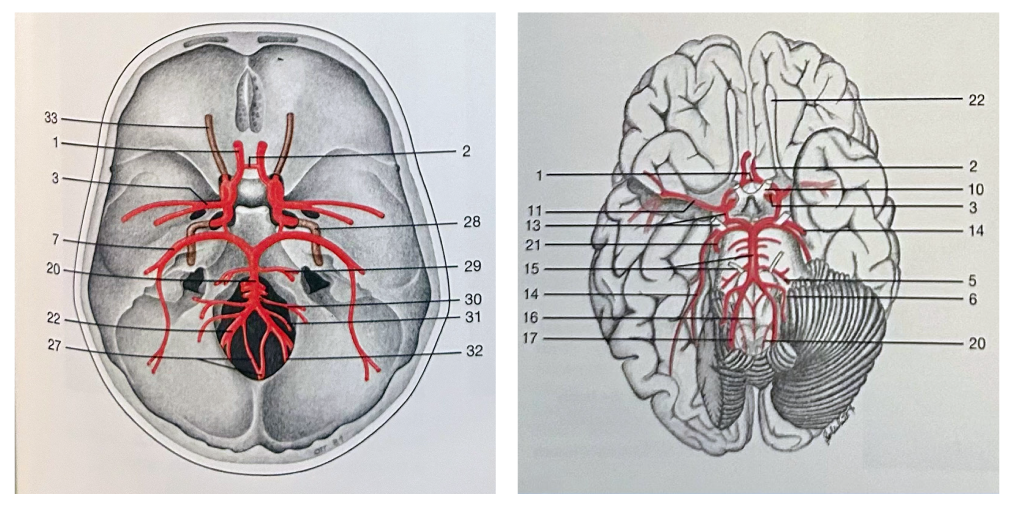
Color Atlas of Anatomy, 3rd Ed. (Rohen / Yokoshi)
The cerebral arterial circle had been a bit of an abstraction until this day in the lab and my subsequent referencing of the images included here. Below you can see two schematics of the arterial circle. The circle is formed by two arteries that enter the cranium; the internal carotid and the common vertebral artery. These two arteries are the source of oxygenated blood for all of the activities of your brain! In a minute we will trace their pathway from the neck into the cranium, but first let’s unpack the circle just a little.
We don’t need to obsess about the names of each little branch but it’s cool to map it out a little bit. The internal carotid artery provides most of the arterial flow for the anterior portion of the brain. The vertebral artery then accounts for most of the arterial flow for the posterior portion of the brain. Cool! The two major branches are then knitted together by little branchlets and that completes the circle!
One of the things this achieves is creating some redundancy in the system in case there is disruption to the flow due to injury or arterial blockage. Also, cool!
Anatomical Footnote: It is interesting that the cerebral arterial circle also surrounds the stalk of the pituitary gland. I could go on and on about the convergence of structures in this part of the cranium; the spheno-basilar joint (jctn of sphenoid and occiput), the temporal bones hugging up the sphenoid, the tentorium arching along the petrous ridge and extending two little feet onto the sphenoid, the arterial circle, the pituitary gland, and the optic chiasma (where the optic nerves merge into a bulb-like structure and then criss-cross and extend into the occipital lobes).
Your Cranium is linked in Meaningful Ways to Your Neck
Where do these arteries come from? The best question. Gil uses a metaphor of a tree to help us visualize the weirdness that is vascular nomenclature. I find it super helpful to imagine a tree that just sends off more and more branches. Now, we would never name every individual branch on our favorite tree. But… that’s what we have in regional anatomy.
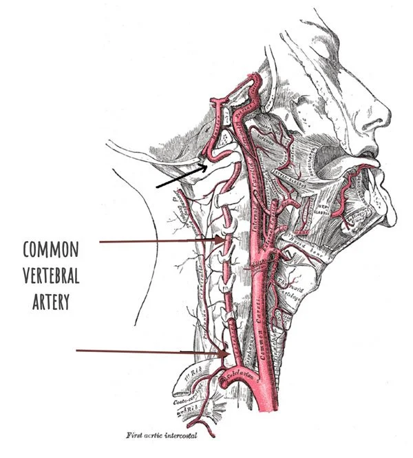
Common Vertebral Artery (Gray’s Anatomy)
Let’s follow inferiorly and see that this arterial circle is just formed of smaller branches off bigger branches. Just like our tree. So, we have climbed the arterial tree from the heart up two different branches of the neck and into the cranium.
The vertebral artery runs along the sides of your cervical spine and enters the cranium through the foramen magnum. It forms two difficult right angle turns to get there. I hypothesize that this area is susceptible to myofascial tension and this could potentially dampen the arterial flow into the cranium. The vertebral artery pathway is responsible for providing arterial circulation to the posterior parts of our brains.
The internal carotid artery runs along the anterior-lateral portion of the neck. The internal carotid is giving arterial blood to the anterior portion of the brain. We can imagine it tucked a bit behind the SCM and within the carotid sheath. The carotid sheath also contains the internal jugular vein and the vagus nerve. On dissection, the sheath almost seems to be embedded within the myofascia of the posterior SCM and the hyoids.
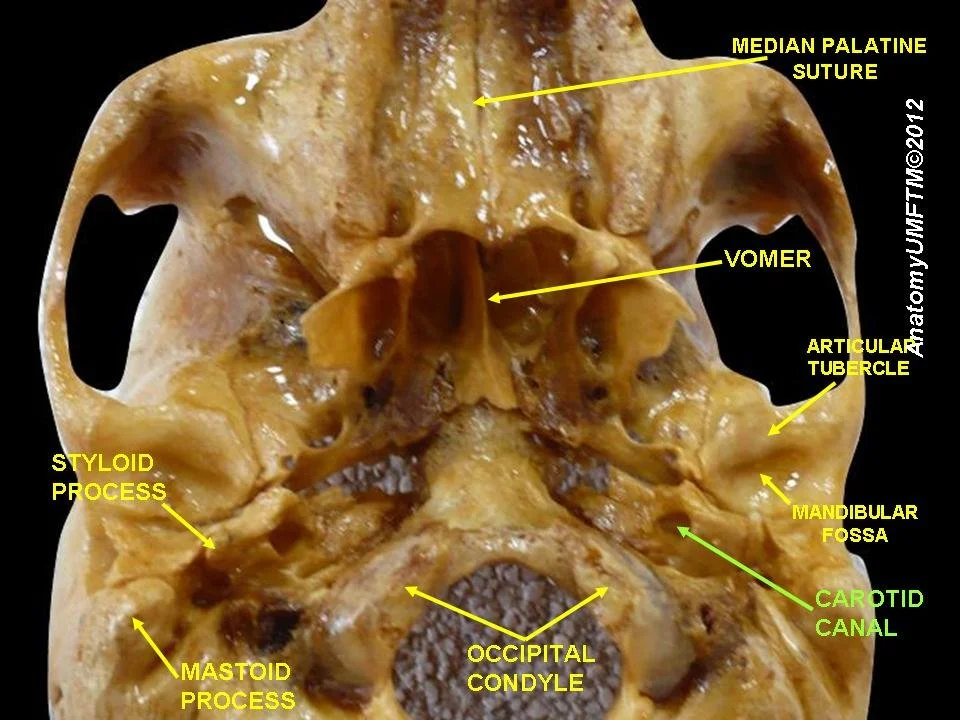
Carotid Canal, 2012 (Victor Babes University of Pharmacy and Medicine)
To enter the cranium, the internal carotid artery passes through a hole in the temporal bone! It’s called the carotid canal.
This is just anterior to the jugular foramen where three major cranial nerves exit the cranium; vagus, glossopharyngeal and spinal accessory nerves. This portion of the temporal bone is wedged in between the basilar portion of the occiput and the sphenoid. These are interesting proximities. Remember, the jugular foramen is formed by little divots carved out of the temporal bones and the occiput, making a “hole” for those nerves.
Clinical Relevance
If we imagine that one of the potential effects of good myofascial work combined with good CST is that we are helping improve overall fluid circulation then it is possible that we are supporting increased arterial flow from the cervical arterial tree into the cranium.
This is where this becomes relevant when treating folks dealing with POTTS and/or dysautonomia. I have seen recent research that points to cervical myofascial tension potentially contributing to the symptoms associated with POTTS/Dysautonomia. These relationships are also potentially useful when thinking about post-traumatic injuries to the cervical region and/or the head. And so much more…
As we follow tissue continuities via a holistic and curious approach to our anatomy studies we break down the unnecessary conceptual barriers created by the regional anatomy approach and enter into a way of study that is more coherent with how the body experiences itself. As I said earlier, I believe bodies can tell when our imagination, our cognitive relationship with the work and our hands reflect a model that the body can relate to. I believe this way of approaching our work creates space for bodies to express themselves because there is an open-minded, curious attitude within the hands and mind-state of the provider. If the provider is working with an attitude of certainty this can override curiosity. Weirdly, the more I internalize images of the body, the more curious I become about it, not the more certain I am. When we approach mystery it has the paradoxical effect of becoming more mysterious!
If you think this approach sounds interesting then you should check out our CST Foundations series and or other offerings. They all step from just this type of thinking.
Endnotes
1. Cleveland Clinic website, accessed on 8/31/2026. https://my.clevelandclinic.org/health/body/circle-of-willis
Recent Posts